Polarization-sensitive optical coherence tomography enables noninvasive whole-donor-liver viability assessment
By
Qinggong Tang
Summary
This article presents a research study where Yan et al. applied polarization-sensitive optical coherence tomography (PS-OCT) to noninvasively image entire donor liver surfaces for viability assessment. The technique addresses the critical shortage of viable donor livers by enabling comprehensive whole-organ evaluation, overcoming the limitations of traditional biopsy sampling and histopathology which cannot detect spatial heterogeneity of liver pathology. The researchers developed imaging parameters and machine learning methods to evaluate steatosis, fibrosis, and inflammation, potentially broadening the classification of viable livers for transplantation — especially important as marginal livers from extended criteria donors are increasingly used.
Source
 Twitter / XPolarization-sensitive optical coherence tomography enables noninvasive whole-donor-liver viability assessmentscim.ag
Key quotes
· 3 pulledLiver transplantation is critically limited by availability of donor organs of sufficient quality.
Current viability assessment by biopsy sampling and histopathology is inherently limited in detecting spatial heterogeneity of liver pathology.
Techniques for comprehensive whole-organ evaluation could broaden viable liver classification.
You might also wanna read


Study evaluates procurement biopsy and machine perfusion for kidney transplant outcome prediction using OPTN database
This retrospective cohort study analyzed data from the Organ Procurement and Transplantation Network database (2014-2022) to evaluate how pr

International multidisciplinary consensus standardizes laparoscopic liver biopsy practices
An international multidisciplinary panel of 45 experts from 6 continents developed a consensus statement on laparoscopic liver biopsy (LLB).
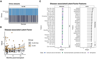
Multi-omics biomarkers in lung fluid predict chronic lung allograft dysfunction after transplantation
This prospective cohort study investigates multi-omics biomarkers of endothelial dysregulation that precede chronic lung allograft dysfuncti
 plos.io·10d ago
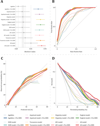
Plasma proteomic signatures enable risk prediction of early retinal neurodegeneration in type 2 diabetes
This multi-cohort prospective observational study by Wei Wang and colleagues investigates circulating plasma protein signatures associated w
plos.io·26d ago
MIT Develops Noninvasive Glucose Monitoring Device Using Raman Spectroscopy
MIT researchers have developed a noninvasive device using Raman spectroscopy to measure blood glucose levels without finger pricks. The shoe
 news.mit.edu·6mo ago
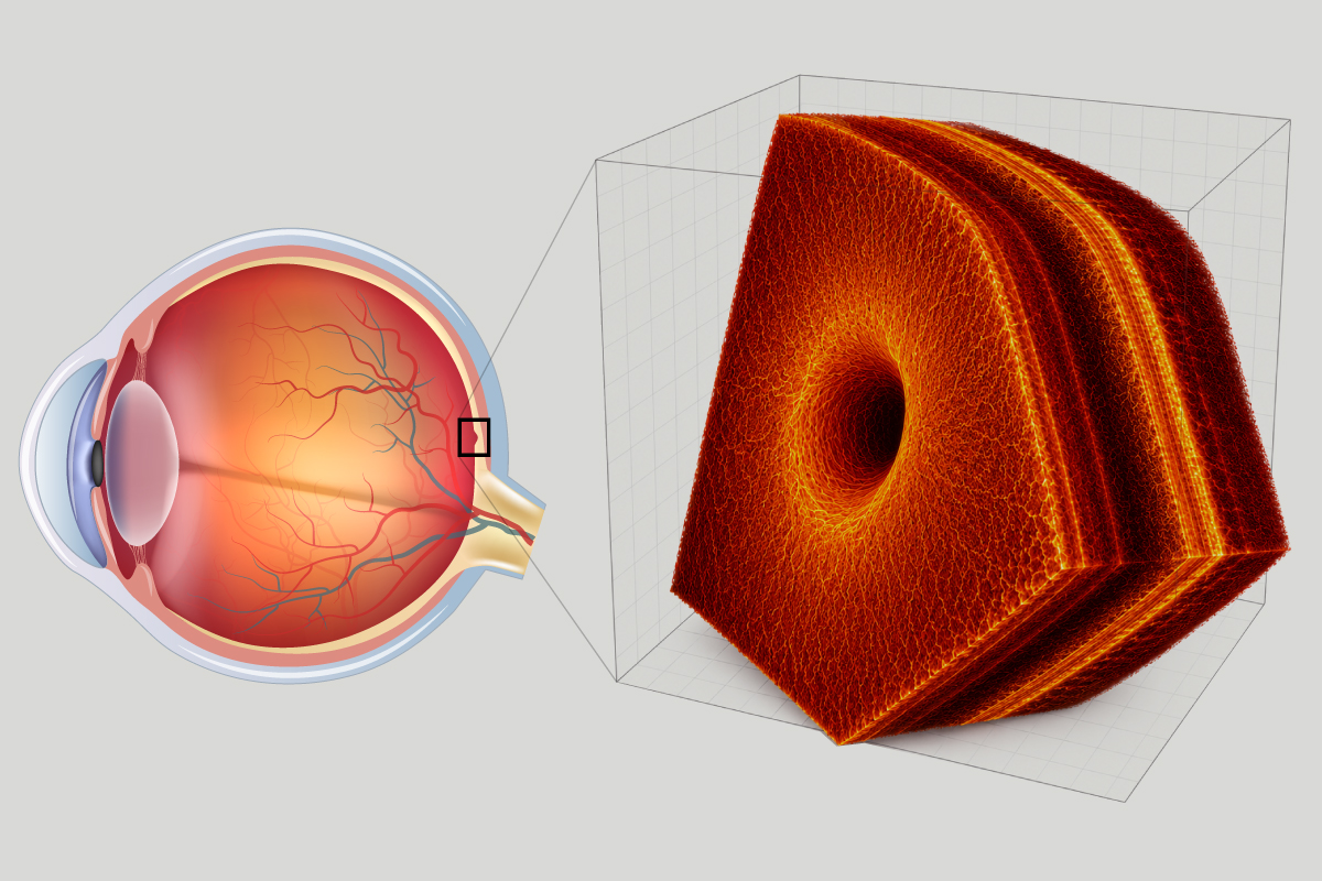
WashU Medicine researchers develop AI system to interpret 3D retinal scans for faster disease diagnosis
Researchers at WashU Medicine have developed an experimental AI system that interprets 3D images of the eye's retina to help diagnose retina
 medicine.washu.edu·26d ago
Comments
Sign in to join the conversation.
No comments yet. Be the first.